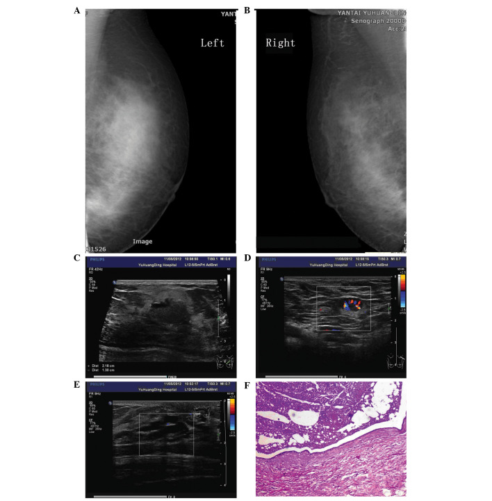
Figure 1.
(A) Left and (B) right mammography revealing thickened breast tissue, without an observable mass, and sand-like calcification in the bilateral breasts. (C) Ultrasound showing the largest mass measuring 2.2×1.3 cm located in the upper inner quadrant of the left breast, with a clear boundary and uneven echo. (D) An enlarged lymph node with a rich blood flow signal around the nodule in the left axilla. (E) A duct of the bilateral breasts exhibiting cystic ectasia. (F) Pathological sample showing breast duct dilatation.

