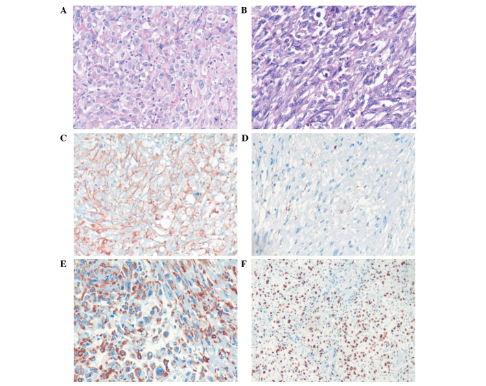
Figure 2.
Histological analysis of the resected tumor specimen. (A) Carcinomatous component base. The tumor cells were irregular and pleomorphic, which can be observed in karyokinesis and necrosis tissues (HE stain; magnification, ×200). (B) Sarcomatoid component base. The morphology of the tumor cells is varied, with the main form being immature and round or spindle-shaped cells. Certain cells possessed an indistinct cell boundary (HE stain; magnification, ×200). Immunohistochemical staining of the resected tumor specimen demonstrating positive expression of (C) creatine kinase (++), (D) epithelial membrane antigen (+), (E) vimentin (++) and (F) Ki-67 (++; 90%). HE, hematoxylin and eosin; (+), weakly positive staining (3–24% cells positively stained); (++), strongly positive staining (25–49% cells positively stained).

