
Figure 3.
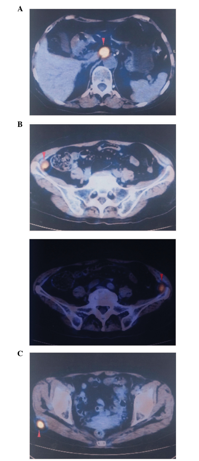
Positron emission tomography-computed tomography scan revealing the initial metastatic lesions in the (A) paraaortic lymph node (SUV value, 9.1), (B) bilateral left and right iliac fossa (SUV value, 4.7) and (C) right gluteal region (SUV value, 7.1). SUV, standardized uptake value.