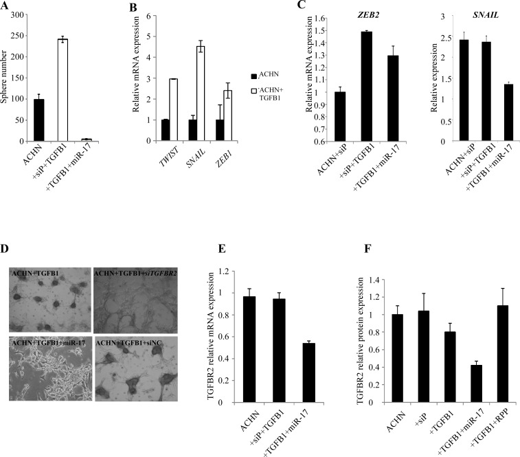
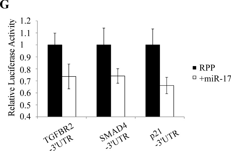
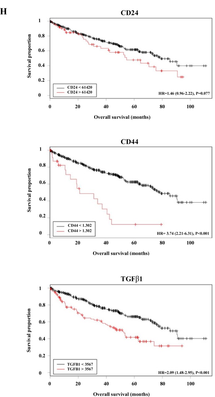
Figure 7. TGFβ signaling is involved in RCC formation and is regulated by miR-17.
(A) TGFβ1 treatment induced sphere formation in ACHN cells. Parallel miR-17 transfection interfered with sphere formation. (B) TGFβ1 treated ACHN cells show increased expression of mesenchymal markers. (C) miR-17 transfection of TGFβ1 treated ACHN cells leads to decreased expression of ZEB2 and SNAIL EMT markers. (D) Silencing of TGFBR2 by siRNA or by miR-17 tra nsfection inhibited RCC sphere formation. miR-17 transfection leads to decreased TGFBR2 expression at mRNA (E) and protein level (F). (F) Quantification of Western blot detecting TGFBR2 expression. (G) CAKI-1 cells were co-transfected with the luciferase constructs containing the 3′ UTRs of TGFBR2, SMAD4 or p21 and miR-17. Decreased luciferase activity indicates that TGFBR2 is a possible direct target of miR-17. (H) Analysis of TCGA database revealed a significant increase of CD24, CD44 and TGFβ1 expression in ccRCC patients with poor survival.


