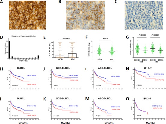
Figure 1. Expression and prognostic significance of CXCR4 in DLBCL.

(A-C) Representative CXCR4 immunohistochemistry staining (showing 100%, 60%, 0% CXCR4 cell surface expression in DLBCL cells). (D) Histogram of CXCR4 expression frequency distribution in the DLBCL study cohort. X-axis, percentage of immunopositive cells in tumors; Y-axis, numbers of DLBCL patients. (E-F) ABC-DLBCL compared to GCB-DLBCL had increased CXCR4 cell surface expression, but did not differ in CXCL12 mRNA significantly. (G) CXCR4 cell surface expression correlated with decreased CXCL12 mRNA levels, both in GCB- and ABC-DLBCL. (H-I) CXCR4 expression correlated with significantly poorer OS and PFS in the overall DLBCL cohort. (J-K) CXCR4 expression correlated with significantly poorer PFS (but not OS) in GCB-DLBCL. (L-M) CXCR4 expression correlated with significantly poorer OS (but not PFS) in ABC-DLBCL. (N-O) CXCR4 expression correlated with significantly poorer survival in DLBCL patients with a low IPI, but not in DLBCL patients with a high IPI.
