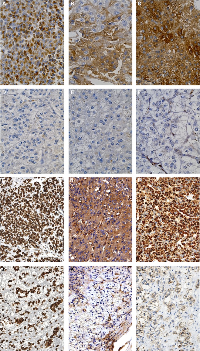
Fig. 5. Immunohistochemical detection of the six proteins differentially expressed in ACC and normal adrenal.

Representative images of immunohistochemistry performed on tumor specimens (n=4) and normal adrenals (n=3) revealed marked positivity to ALDH6A1 (A), Transferrin (B), Fascin-1 (C), CAP-1 (H) and Ferredoxin reductase (I) in the cytosol of almost all tumor cells in the field, compared to no positivity in normal adrenal, respectively (D-F, K-L). Nuclear positivity to Lamin A/C was diffused in tumor cells (G) and less expressed in normal adrenal cortex (J).
