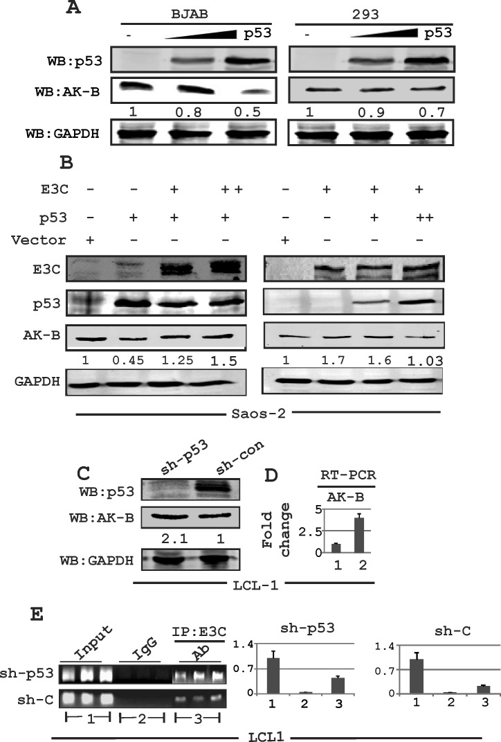
Fig 2. Expression of AK-B in EBNA3C positive and EBV transformed LCLs.
(A) Evaluating the levels of AK-B with dose dependent increase of p53 in BJAB and HEK-293T cells. (B) Evaluation of AK-B in a dose dependent manner in EBNA3C and p53 in Saos-2 (p53−/−) cells. (C, D) Determination of AK-B and EBNA3C at the protein and transcript levels in sh-p53 and sh-control in LCL1 stable cell line. Here 1 and 2 denoted to sh-control and sh-p53 in LCL1 cells. (E) In ChIP assay: stable sh-p53 and sh-control clones LCL1 cells were immunoprecipitated with A10 antibody, followed by real time PCR with AK-B ChIP primers. The amplified PCR products were fractionated on agarose gels. Here 1, 2 and 3 denoted to input, control IgG and IP:E3C group respectively.

