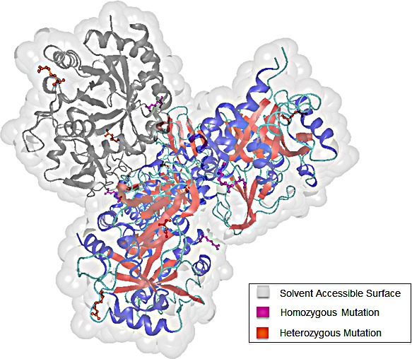
Figure 7. 3D structure of chitinase-3-like protein 1 (CHI3L1).

The four chains of CHI3L1 are colored according to their secondary structure elements. To highlight the distribution of the detected mutations within one chain, chain C is colored in grey. We differentiate between homozygous (pink) and heterozygous (orange) mutations.
