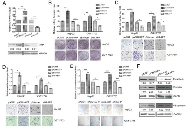
Figure 4. AFP contributes to the malignant phenotypes of HCC.

(A) The qRT-PCR and western blot were used to test the efficiency of pA3M1/AFP and pSilenser2.1-neo/shR-AFP. (B) Colony formation assays were performed to test the influence of AFP on the proliferation of HepG2 and QGY-7703 cells. (C, D) Transwell migration and invasion assays were performed to detect the effect of AFP on the migration (C) and invasion (D) of HepG2 and QGY-7703 cells. (E) The effect of AFP on the VM in the HepG2 and QGY-7703 cells by means of three-dimensional Matrigel culture. (F) The influence of AFP on the protein levels of EMT-associated molecules (E-cadherin and vimentin) and of a key VM molecule (VE-cadherin) was determined by western blotting. To detect the level of VE-cadherin, the QGY-7703 cells were transfected for 60 h before the protein was harvested. *p<0.05, **p<0.01, ***p<0.001. All error bars indicate the means±SDs. All experiments were repeated at least three times.
