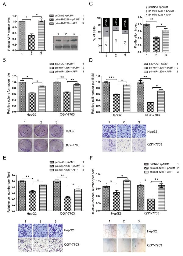
Figure 5. The ectopic expression of AFP counteracts the inhibition of the aggressive malignance induced by miR-1236.

(A) HepG2 and QGY-7703 cells were cotransfected with pcDNA3/pri-miR-1236 and pA3M1/AFP without its 3′UTR or the control vector and then western bolt assay was used to test the restoration of AFP protein by pA3M1/AFP in the presence of miR-1236 (Right). The quantification of the bars are shown on the left. (B) The transfected cells were submitted to colony formation assays to test the proliferation of HCC cells. (C) Cell cycle progression of the transfected cells was analyzed by flow cytometry. The left side of the chart shows populations of cells in the different phases of the cell cycle, and the right side shows the proliferation index. (D-F) Transwell migration/invasion assays and three-dimensional Matrigel culture to test the cells' abilities to migrate (D), invade (E) and undergo VM (F). *p<0.05, **p<0.01, ***p<0.001. All error bars indicate the means±SDs. All experiments were repeated at least three times.
