
Figure 8. NIBP maintains cancer cell survival.
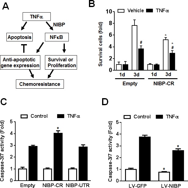
(A) Schematic representation of the cancer cell NIBP/NFκB-dependent regulation of chemoresistance. (B) TNFα-induced cell death in HCT116 cells with NIBP knockdown was examined. An equal number of cells (10,000 cells/well) were seeded in 96-well plates. After 24 h, cells were treated with or without TNFα (10 ng/ml) for 1 and 3 d and the viable cell number was determined by trypan blue staining and hemocytometry. The data represent fold change related to the corresponding vehicle control at 1 d. (C) NIBP shRNA knockdown sensitized TNFα-induced apoptosis in HCT116 cells. Equal numbers of cells stably engineered with indicated shRNA were cultured in a 96-well plate (5,000 cells per well in quadruplicate) and treated with TNFα (10 ng/ml) for 24 h before Caspase-Glo® 3/7 assay was performed. (D) NIBP overexpression inhibited constitutive and TNFα-induced apoptosis in HCT116 cells. Cells were infected with indicated lentivirus (LV) and the third passage cells (5,000 cells per well in quadruplicate) were treated with TNF (10 ng/ml) for 24 h before Caspase-Glo® 3/7 assay was performed. * P<0.05 indicates a significant difference compared with the corresponding empty lentiviral vector.