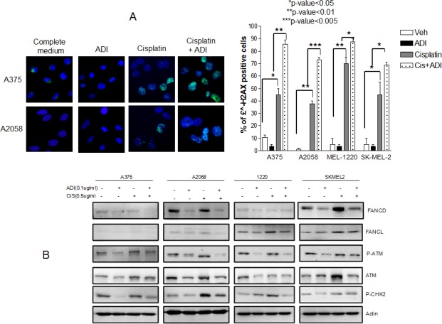
Fig. 4. Increased DNA damage in 4 cell lines when treated with combination treatment.

(A) Immunofluorescence of γH2AX foci after treatment with cisplatin at 0.5 μg/ml for 48 hr in normal media and in ADI-PEG20 treated media. The γH2AX foci increased in all 4 cell lines with cisplatin alone (p≤0.05 in A375 and SkMel2, p≤0.01 in A2058 and Mel1220) which was expected since cisplatin is known to cause DNA damage. Upon arginine deprivation alone, there were no changes in γH2AX foci compared to control; however, the γH2AX foci increased significantly on combination treatment in all 4 cell lines, which indicates that in the absence of arginine, cisplatin treatment results in more damage (p<0.01 in A-375, p<0.005 in A-2058, p<0.05 in Mel-1220 and Sk-Mel2). (B) Illustrates that increase in DNA damage is secondary to decrease in DNA repair process. Ubiquitinated FANCD, p-ATM, ATM, and p-CHK2, which increased in response to cisplatin treatment to initiate the repair process, showed a decrease upon combination treatment. The amount of FANCL also slightly decreased upon combination treatment in 3 cell lines. A375 possess very low amounts of FANCL and so differences could not be visualized.
