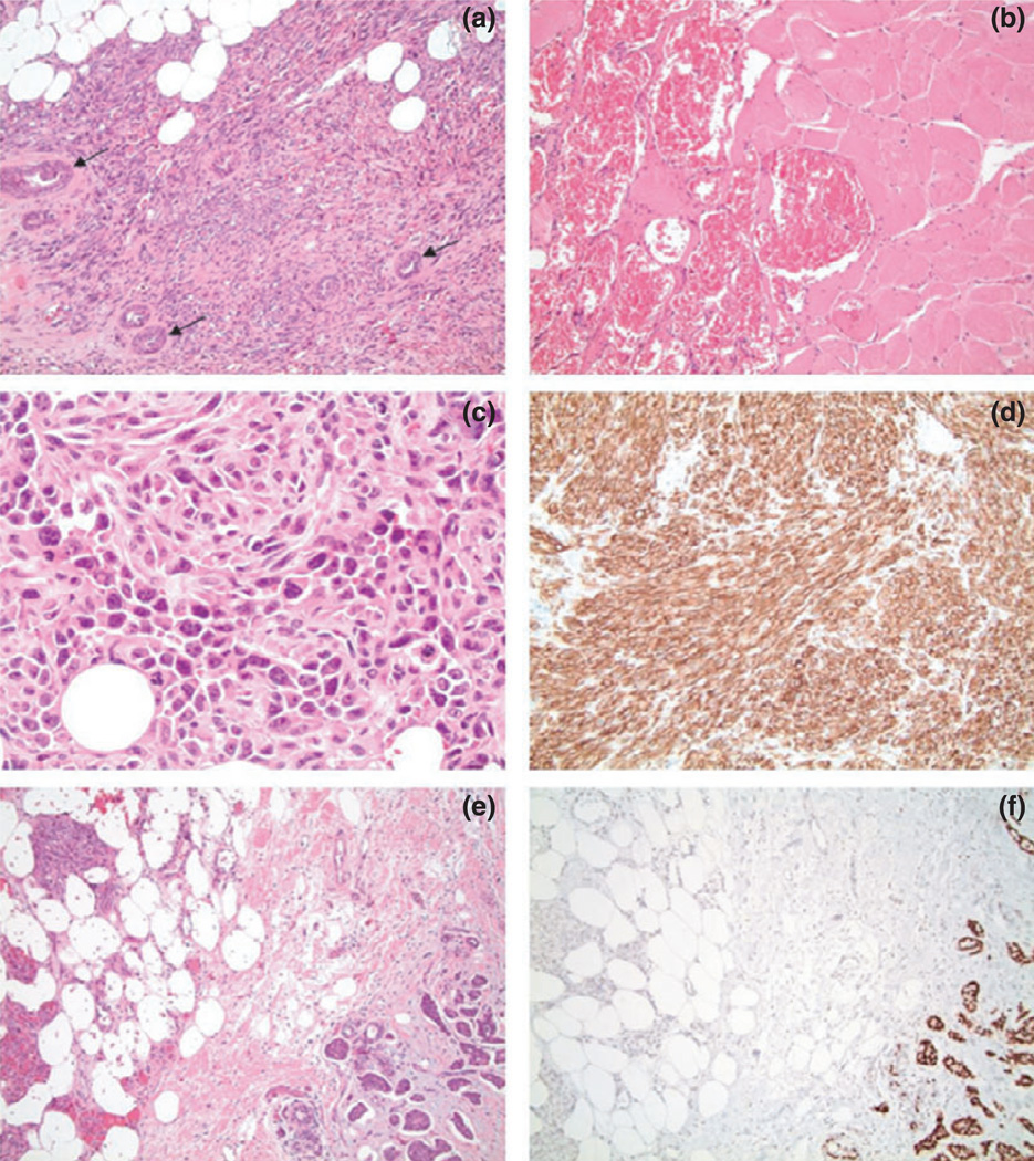
Figure 3.
(a) Hematoxylin and Eosin (H&E) stained slides at of the surgical specimen depicting angiosarcoma with residual entrapped breast ducts (arrows) (10× magnification). (b) H&E at revealing large vascular structures filled with blood invading skeletal muscle (20× magnification). (c) H&E of a solid area of the angiosarcoma depicting high-grade features including nuclear atypia and mitotic figures (40× magnification). (d) CD34 immunostain of the angiosarcoma with strong expression, corroborating the diagnosis of angiosarcoma (20× magnification). (e) H&E demonstrating collision of angiosarcoma (left) and infiltrating ductal carcinoma (right) (10× magnification). (f) Estrogen receptor (ER) immunostain corresponding to the H&E in the prior image showing strong nuclear positivity in the infiltrating ductal carcinoma (right) while the angiosarcoma (left) is negative (10× magnification).

