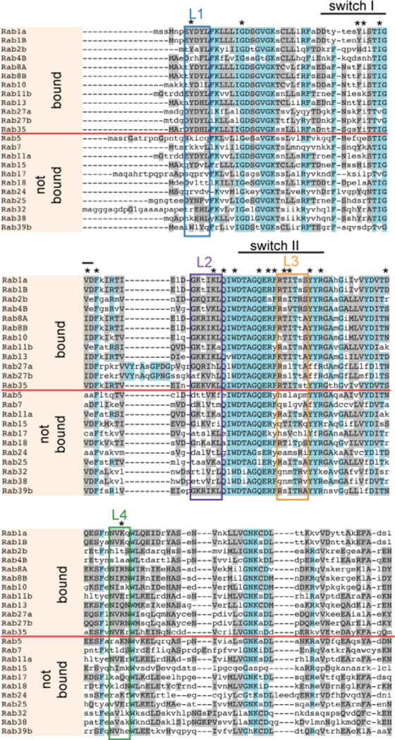
Figure 4.
Conserved regions within Rab proteins targeted by LidA. Sequence alignment of Rab GTPases was performed using the MUSCLE server (http://www.bioinformatics.nl/tools/muscle.html). Rabs that interact with LidA are separated by a red line from those that do not interact. Regions of high homology are shown in magenta; regions of limited homology are shown in grey. Clusters of enhanced conservation are labeled by boxes. * indicates amino acid residues known to be involved in LidA-Rab1 binding (23).

