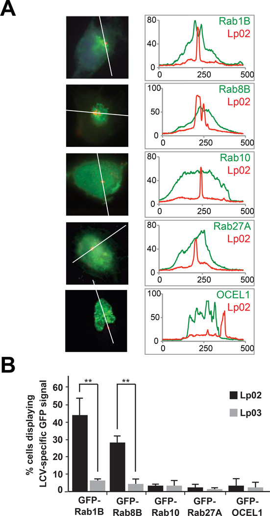
Figure 7.
Localization of host targets to the LCV. (A) CHO-FcγRII cells were transfected with constructs encoding the indicated GFP-tagged Rab proteins or OCEL1, and challenged with L. pneumophila Lp02 (wild-type) or Lp03 (T4SS mutant). Intracellular bacteria (red) were detected with anti-L. pneumophila antibody. Scale bar, 1µm. Line scans (right panels) denote pixel intensity of red and green fluorescent signals along the indicated line. (B) Quantification of (A) showing the percentage of cells displaying an LCV-specific GFP signal. Data are mean ± SD (error bars) for three independent experiments. **P < 0.01 (two-tailed t-test).

