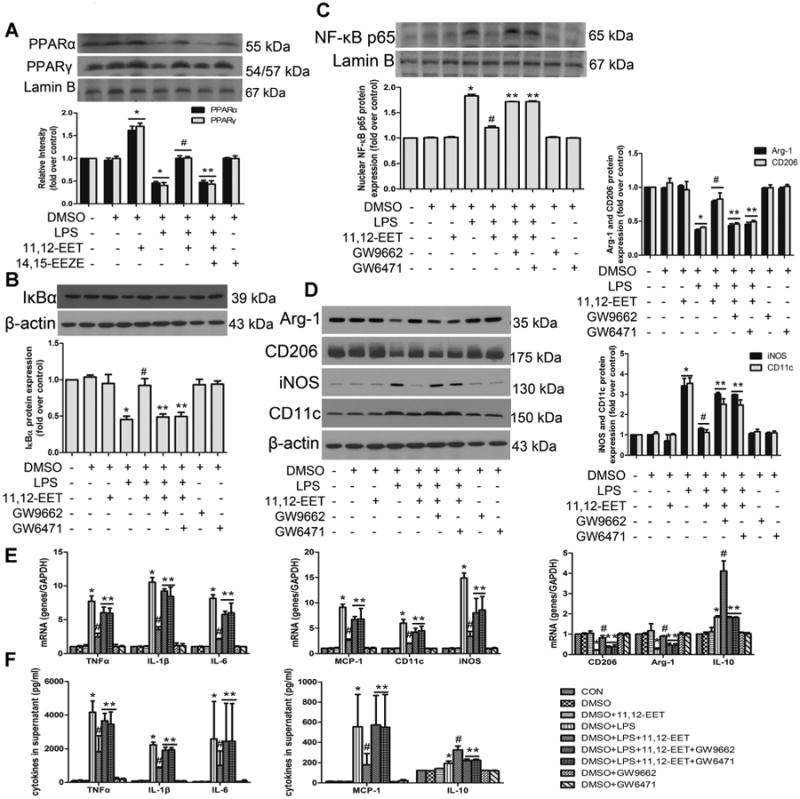
Fig 3.

11,12-EET mediated M1 to M2 phenotype polarization and anti-inflammatory effects are mediated by PPARα/β activation in primary cells. A: Representative immunoblots and quantitation of peroxisome proliferator-activated receptor α and γ (PPARα and PPARγ) expression in nuclear following 11,12-EET treatment in the presence or absence of LPS in primary cells. Representative immunoblots and quantitation of NF-κB signaling pathway (B, C) and M1, M2 markers (D). Administration of PPAR antagonists (GW9662/GW6471) significantly abolished EET effects on LPS-induced M1 macrophage polarization. E: Relative mRNA expression of inflammatory genes (TNFα, IL-1β, IL-6, MCP-1, IL-10) or phenotype molecules (CD11c, iNOS, CD206, Arg-1) were determined after LPS stimulation for 3 h. F: TNFα, IL-1β, IL-6, MCP-1, and IL-10 were assessed by ELISA after LPS stimulation for 12h. Data are shown as mean ±SEM from three independent experiments. *P< 0.05 versus control; #P<0.05 versus DMSO + LPS; **P< 0.05 versus DMSO + LPS + 11,12-EET.
