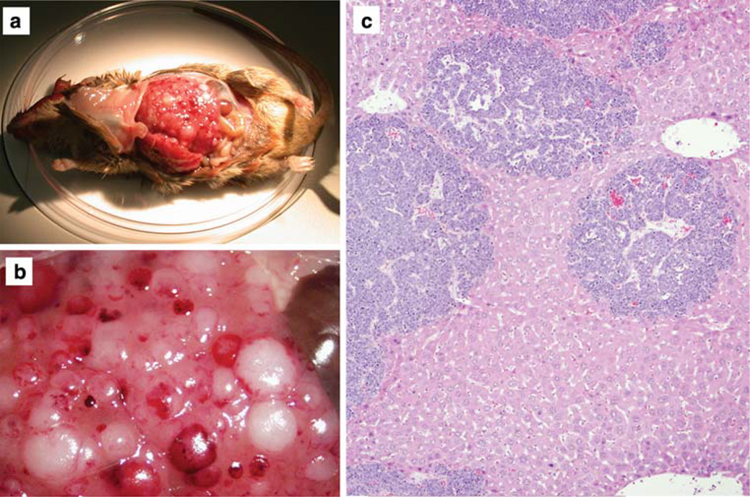
Fig. 2.
Liver metastasis in a Tyr-TAg/Id2+/− mouse. a Low magnification external photograph. b High magnification external photograph, showing multiple foci of metastatic lesions on the liver surface. c Histopathologic photomicrograph showing multiple dark blue foci representing intrahepatic metastases. Original magnification 40×

