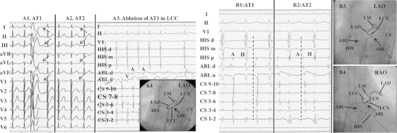
Figure 4.

Surface ECG features and catheter ablation of 2 anterior atrial septum-atrial tachycardias (ATs) from the noncoronary cusp (NCC) and aortic mitral junction (AMJ) in 1 patient. A, P-wave morphological features on a 12-lead ECG during 2 ATs and termination of AT1 in the left coronary cusp (LCC). A1 and A2, Surface ECG of AT1 and AT2. The cycle length of AT1 and AT2 was 470 and 510 ms, respectively. A3, Ablation at the LCC could temporarily terminate AT1. A4, Catheter location at the LCC in the LAO projection. The white and black dotted lines represent the schematic anatomy of the left coronary artery and the LCC, respectively, according to the coronary angiogram. B, The intracardiac electrograms and the fluoroscopic images at the successful ablation sites of 2 ATs. B1, AT1 was eliminated at the aortic mitral junction (AMJ). B2, AT2 was eliminated at the NCC. Of note, the different atrial activation sequences between AT1 and AT2 as the coronary sinus (CS) mapping catheter was inserted further distally into the CS. B3 and B4, The fluoroscopic images of successful ablation sites of 2 ATs. Of note is the anatomic vicinity between the LCC and AMJ. Asterisk, ablation site at the LCC; solid arrow, ablation site at the NCC; and dotted arrow, ablation site at the AMJ. ABL indicates ablation catheter; LAD, left anterior descending branch; LCX, left circumflex branch; and LM, left main coronary artery.
