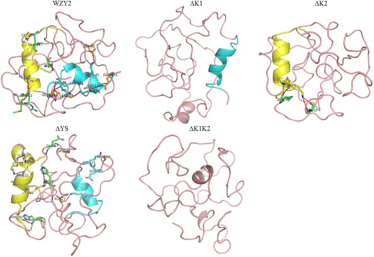
FIGURE 2.
Three-dimensional structure prediction of WZY2 and its truncated derivative polypeptides. The K1-segment is shown in yellow, and the K2-segment is in blue. The amino acid residues involved in the helices-protein interaction are shown in yellow (in the K1-segment), in green (with the K1-segment), in blue (in the K2-segment), and in orange (with the K2-segment).

