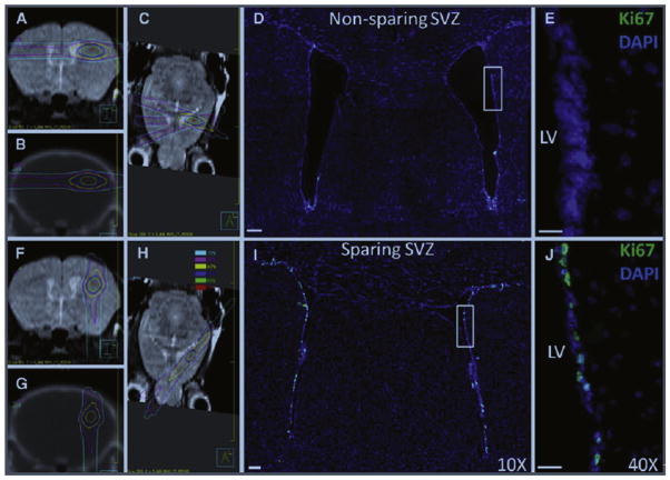
Figure 5.
Mouse radiation treatment plans (left) and microscopy images (right) for the non–NPC-sparing (top) and NPC-sparing (bottom) radiotherapy plans from Redmond et al.43 Left side MR and CT images from the mouse radiation treatment plans showing the radiation dose distribution for the non–NPC-sparing (A–C) and NPC-sparing radiation treatment plans (F–H). It can be noted that for the non–NPC-sparing plan, the region of the SVZ of the ipsilateral lateral ventricle receives a high radiation dose, whereas this region is effectively spared in the NPC-sparing plan. Scans taken are as follows: coronal MRI (A and F), coronal CT (B and G), and axial MRI (C and H). Dose values are shown in the legend. Right side coronal sections showing Ki-67 stains (green) in the SVZ of the lateral ventricles following non–NPC-sparing RT (D and E) and NPC-sparing RT (I and J). Ki-67 is a marker of cellular proliferation and is used in this model as a potential indicator of NPCs. Costaining is performed using DAPI (blue). Images (D and I) were taken with a 109 objective lens and the images (E and J) with 409 objective lens. CT, computed tomography; DAPI, 4′,6-diamidino-2-phenylindole; MRI, magnetic resonance imaging; LV, left ventricle.

