Fig. 6.

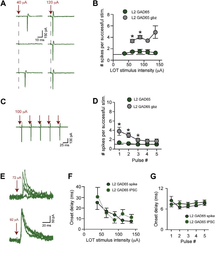
Well-timed inhibition limits the spike response of L2 GAD65-eGFP cells. (A) Cell-attached recording of spike activity in an L2 GAD65-eGFP cell. Responses are shown to moderate intensity stimuli (40 μA) that were perithreshold for generation of spikes and, also, to high-intensity stimuli (120 μA). The number of evoked spikes when there were spikes remained constant, at one. (B) The mean number of spikes evoked during successful trials was invariant ranging between 1 and 2 across LOT stimulus intensity. Gabazine (10 μM; gbz) increased the number of evoked spikes. N values for control data points ranged between 4 and 17. (C) Spike activity recorded in an L2 GAD65-eGFP cell in response to a 20-Hz stimulus train applied to LOT (100 μA). (D) The mean number of evoked spikes during successful trials as a function of stimulus number during a 20-Hz train in L2 GAD65-eGFP cells. LOT stimulus intensity was 100 μA for all recordings. (E) Outward-going IPSCs (at Vhold = −7 mV) in an L2 GAD65-eGFP cell in response to LOT stimulation at two stimulus intensities. Five trials are shown for each intensity. (F) The onset delays for the first spike measured in cell-attached recordings were well-matched to the first IPSC across LOT stimulus intensity. N values for each point ranged from 3 to 15. (G) The onset delays of the first IPSCs recorded in response to 20-Hz stimuli (raw data not shown) were well-matched to the first spikes in L2 GAD65-eGFP cells.
