Fig. 2.
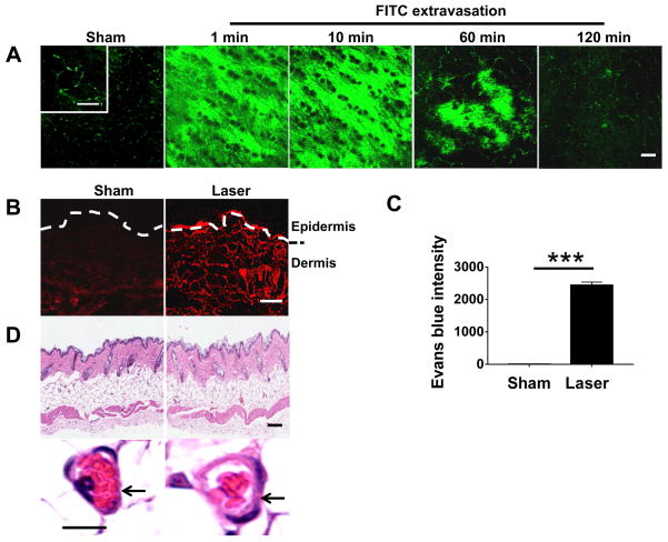
Laser induces extravasation. (A) FITC extravasation occurs rapidly in the skin illuminated by 532 nm NYL laser with a fluence of 0.5 J/cm2. Intravital laser confocal microscopy was used to track FITC signal over time after laser illumination in the skin of mice that had received FITC intravenously. Control skin was shown after illumination with sham light in the same mice. (B) Diffusion of Evans blue dye throughout the dermis after laser illumination. (C) Evans blue intensity increases in upper dermis by more than 1000 times in laser-treated skin as compared with non-laser-treated skin. (D) Histological analysis of control and laser-treated skin. Arrows indicate a capillary vessel. Scale=200 μm in (A), 50 μm in (B), 100 μm in (D, upper) or 5 μm in (D, bottom).

