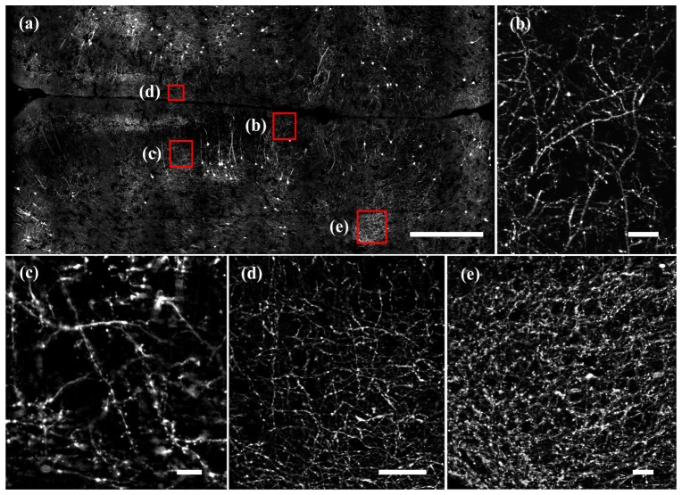
Fig. 6.
Imaging of a large area mouse brain slice. (a) The imaged region is about 1.628 mm wide and 3.328 mm long. Scale bar: 500 μm. (b) (c) (d) (e) Enlarged images of brain regions marked as red box in (a). The enlarged images demonstrate visualization of dendrites spine, buttons, and axon fibers in the whole imaging regions. Scale bar, 20 μm.

