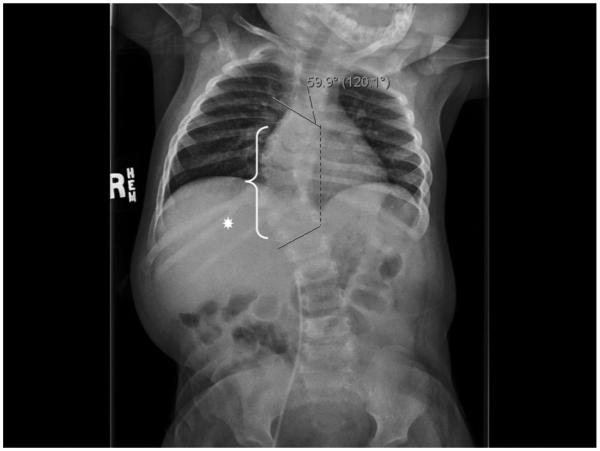
FIG. 1.

Patient plain skeletal film displaying her severe 59.9° thoracic dextroscoliosis. Based on 3-D CT reconstruction (not shown) the dextroscoliosis appears to have its apex at the T10/11 level. Multiple vertebral anomalies were also identified (white bracket), including right hemivertebrae at levels T8 and T11. The right T8 hemivertebra was partially fused to the right aspect of T9. Additionally, there was clefting of vertebral bodies T6 and T9 and fusion of the posterior elements of T11 and T12. There also appeared to be a spina bifida occulta at both the T11 and T12 levels. There are only nine ribs on the left side. Note is made of a bifid right rib at the T11 level (white asterisk).
