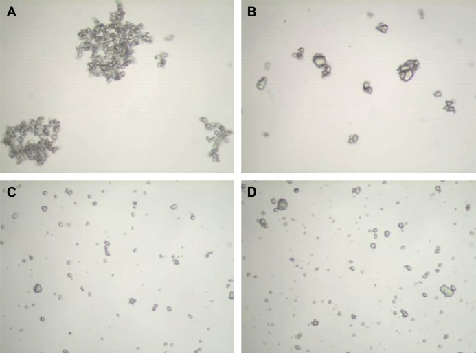
Figure 1.
Representative images of particles of Triesence® (A, C) and Kenalog®-40 (B, D). In (A and B), samples were diluted in buffered salt solution. In (C and D), samples were diluted in a solution containing 1 g/L Triton X-100. Samples were photographed with a Nikon Labophot polarizing light microscope with Clemex Vision Lite 4.0 software. Images were taken at a magnification of 400×.

