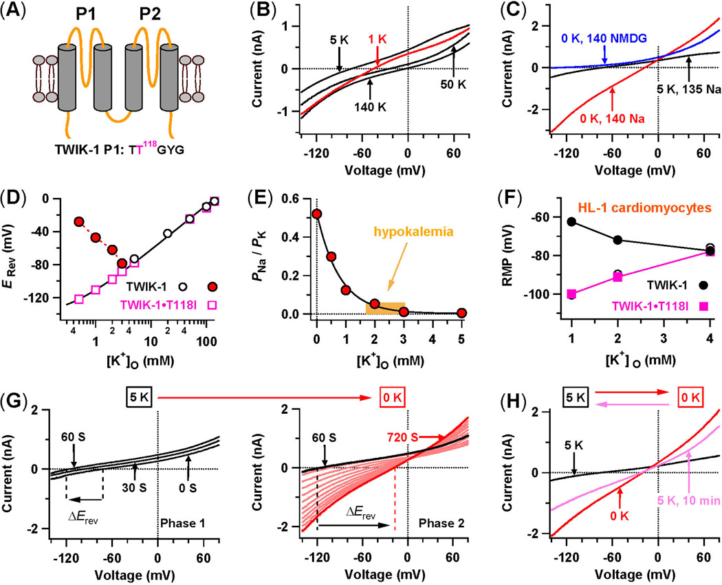
Figure 2. In hypokalemic conditions, TWIK1 K+ channels conduct inward leak Na+ currents and induce cardiac paradoxical depolarization.

(A) TWIK1 topology with four transmembrane domains (TM1 to TM4) and two P-loops (P1 and P2). The sequence of its K+ selectivity sequence TTGYG in P1 is shown. (B) Whole-cell TWIK1 currents in transfected CHO cells in different external K+ concentrations. (C) Direct recordings of inward leak Na+ currents. Whole-cell TWIK1 currents are shown while Na+-based bath solutions with 5 mM K+ are shifted to 0 mM K+, and finally to an NMDG+-based bath solution with 0 mM K+. (D) Reversal potentials (Erev) measured in Na+-based bath solutions with various [K+]o. The continuous line is a fit for open squares with the Goldman-Hodgkin-Katz voltage equation. (E) PNa/PK values in normal and low [K+]o. (F) Resting membrane potentials (RMP) of mouse HL-1 cardiomyocytes with and without over-expression of TWIK1 and TWIK-1•T118I in 1, 2, and 4 mM [K+]o. (G) Two-phase changes of whole-cell TWIK-1 currents are shown while Na+-based bath solutions are changed from 5 mM to 0 mM K+. (H) Whole-cell TWIK1 currents are shown when a Na+-based bath solution is reversibly changed from 5 mM to 0 mM K+, and then back to 5 mM K+ for 10 minutes. (adapted from [13]).
