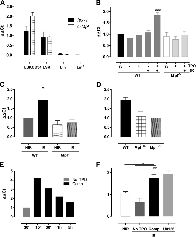
Figure 3.
Iex-1 is induced in HSPCs in response to TPO and IR. (A) Quantitative polymerase chain reaction analysis of Mpl and Iex-1 mRNA levels in different hematopoietic cell populations. Results are means + SEM (n = 3). (B-F) Quantitative polymerase chain reaction analysis of Iex-1 expression in LSK cells from WT, Mpl−/−, and Mpl+/− mice cultured in vitro in complete medium (Comp) or without TPO and irradiated or not (2 Gy). Analysis was performed at 5 hours (B,D) or at various times after IR (E). (F) Cells were incubated with the MEK inhibitor U0126 (10 µM) 30 minutes before being treated as in panel B. Data are means + SEM (n = 4). (C) Iex-1 mRNA expression in LSK cells isolated 5 hours after TBI of WT and Mpl−/− mice. Data are means + SEM (n = 3). All the results are normalized on gapdh expression.

