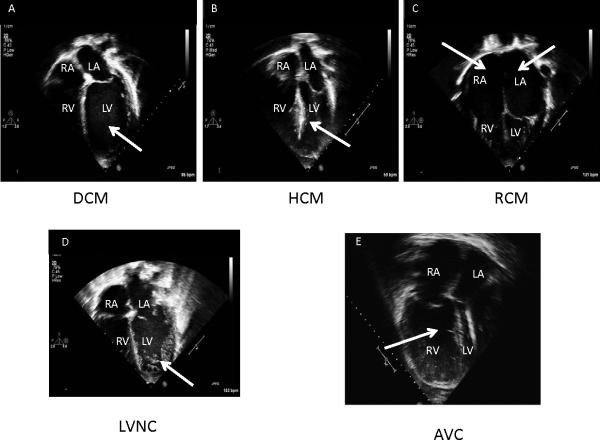
Figure 2. Echocardiographic features of cardiomyopathies.
Panel A. 4-Chamber echocardiographic view of dilated cardiomyopathy (DCM). Note the dilated left ventricle (LV; arrow); Panel B. 4-Chamber echocardiographic view of hypertrophic cardiomyopathy (HCM). Note the thickened mid-portion of the interventricular septum (arrow); Panel C. 4-Chamber echocardiographic view of restrictive cardiomyopathy (RCM). Note the dilated atria (RA, LA; arrows); Panel D. 4-Chamber echocardiographic view of left ventricular noncompaction cardiomyopathy (LVNC). Note the hypertrabaeculation in the LV (arrow); Panel E. 4-Chamber echocardiographic view of arrhythmogenic right ventricular cardiomyopathy (ARVC). Note the dilated, trabeculated right ventricle (RV; arrow); Commonly, an aneurysm of the RV or RV outflow tract can be seen, particularly by MRI.

