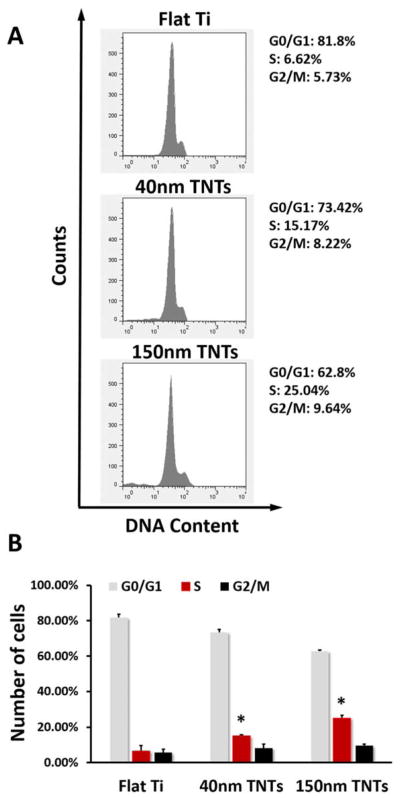
Fig. 3.
(A) Flow cytometry analysis of MC3T3-E1 cell cycle on different substrates after 24h culture, (B) Percentage of MC3T3-E1 cells in G1, S and G2 phases on different substrates. The statistical significance (p < 0.05) after performing t-tests are marked: *, indicates a significant difference between the same phase of the cells growing on different substrates, compared with Flat Ti.

