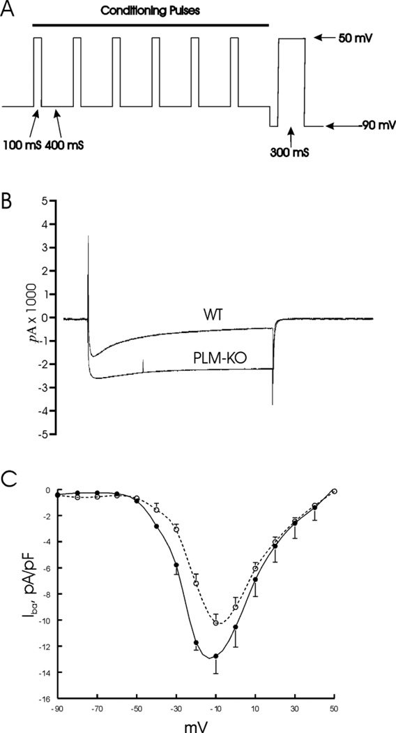
Figure 2.
PLM enhances voltage-dependent inactivation of ICa. ICa was measured in freshly isolated myocytes from WT and KO hearts, using Ba2+ (2 mM) as permeant ion. A. Voltage-clamp protocol: test pulses from −90 to +50 mV (in 10 mV increments) were extended from 60 to 300 ms duration (for simplicity, only one test pulse is shown). B. Representative IBa traces at −10 mV from WT and KO myocytes are shown. Note prolonged time course of IBa in KO compared to WT myocyte. C. I-V curves from WT (○; n=5) and KO myocytes (●; n=5).

