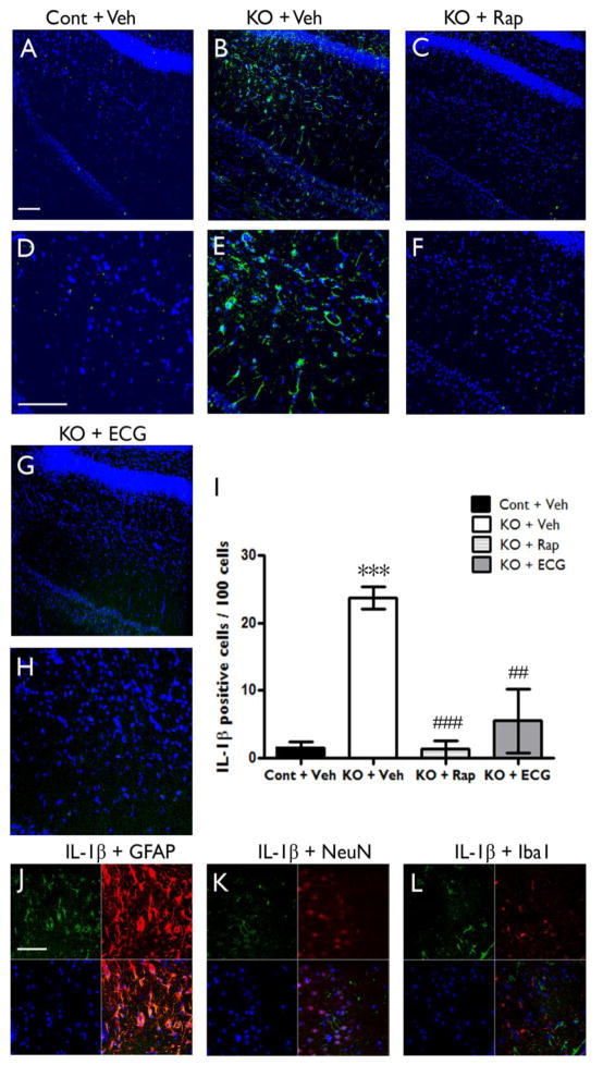
Figure 2. IL-1β protein is upregulated in Tsc1GFAPCKO mice and inhibited by anti-inflammatory treatments.
Protein expression of IL-1β was assessed by immunohistochemistry in Tsc1GFAPCKO and control mice, as well as in rapamycin or ECG treated Tsc1GFAPCKO mice. (A–H) Confocal images of immunohistochemical staining of IL-1β (green) and TO-PRO-3 Iodide (blue). TO-PRO-3 Iodide (blue) was used as an optimal fluorescence dye for nuclear counterstaining. IL-1β protein expression was detected in brain sections of Tsc1GFAPCKO mice (B,E KO + Veh), but not in control mice (A,D Cont + Veh). Rapamycin (C,F KO + Rap) and ECG (G,H KO + ECG) treatment inhibited the IL-1β expression. Scale bars = 100 μm. (I) Quantitative analysis confirmed an increase in IL-1β-positive cells (per 100 TO-PRO-3 positive cells) in vehicle-treated Tsc1GFAPCKO group (KO + Veh) compared with vehicle-treated control group (Cont + Veh). Rapamycin (3 mg/kg/d i.p. for one week) or ECG treatment (12.5 mg/kg/d i.p. for one week) significantly decreased IL-1β-positive cells. ***p<0.05 versus vehicle-treated control mice (n=4–5 mice/group); ## p<0.01, ### p<0.001 versus vehicle-treated Tsc1GFAPCKO mice by two-way ANOVA. (J, K, L) Double label immunofluorescence confocal microscopy for expression of IL-1β protein (green) with GFAP (F, red for astrocytes), NeuN (G, red for neurons) and Iba1 (H, red for microglia) within brain sections of four-week-old Tsc1GFAPCKO mice. All sections were also labeled with TO-PRO-3 Iodide (blue) as an optimal fluorescence dye for nuclear counterstaining. IL-1β was found co-localized with GFAP, but not with NeuN and Iba1. Scale bar = 50 μm. Cont = control, KO = Tsc1GFAPCKO, TO PRO3 = TO-PRO-3 Iodide, ECG = Epicatechin-3-gallate, Rap = Rapamycin.

