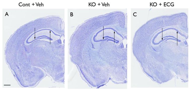
Figure 6. ECG treatment does not prevent neuronal disorganization in Tsc1GFAPCKO mice.

The effect of ECG on neuronal organization was assessed in Tsc1GFAPCKO mice by cresyl violet staining. Compared with control mice (A), vehicle-treated Tsc1GFAPCKO mice (B) exhibited widely dispersed pyramidal cell layers (arrows) in all regions of hippocampus (CA1–CA4). ECG treated Tsc1GFAPCKO mice (C) had a similar pattern as vehicle-treated Tsc1GFAPCKO group (B), with no apparent effect on this neuronal disorganization. Scale bar = 500 μm. Cont = control mice, KO = Tsc1GFAPCKO mice, Veh = vehicle, ECG = Epicatechin-3-gallate.
