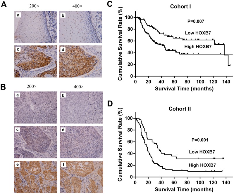
Fig 1. Immunohistochemical detection of HOXB7 in ESCC specimens and Kaplan-Meier survival curve for ESCC patients.
(A) Representative sample of paired normal tissues (a, b) and ESCC (c, d). In the normal esophageal epithelial, HOXB7 expression was mainly limited to the nucleus of the epithelial cells located in the basal and suprabasal layers, whereas in the ESCC, HOXB7-positive cells were broadly observed in the tumor (Case no. 53865). (B) Figure a and b, negative control, with primary antibody replaced by PBS (Case no. 40146). Figure c and d, low expression of HOXB7 in ESCC (Case no. 42873). Figure e and f, high expression of HOXB7 in ESCC (Case no. 36840). (C) Kaplan-Meier survival curve for 177 patients in the cohort I. The median survival time was 45 months for high expression patients, which was significantly shorter than the 137 months for low expression patients (P = 0.007). (D) Kaplan-Meier survival curve for 103 patients in the cohort II. The median survival time was 19 months for high expression patients, which was significantly shorter than the 34 months for low expression patients (P = 0.001).

