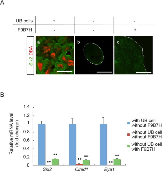
Fig 1. NPC maintenance in aggregates requires UB cells.

(A) Immuno-staining of E11.5 aggregates for NPC marker, Six2 (green), and UB marker, DBA (red). (a) A representative aggregate made from dispersed E11.5 whole embryonic kidney cells and cultured for 7 days. Abundant Six2+ NPC were present surrounding UB structures. (b) A representative aggregate without UB cells reconstituted from E11.5 Hoxb7-Venus mouse embryonic kidneys by manually separating Venus+-UB from the surrounding Venus--mesenchyme cells. No Six2+ NPC were detected after cultured for 7 days. (c) A representative aggregate without UB cells and treated with Fgf9, Bmp7 and heparin (F9B7H) for 7 days in culture. Only a few Six2+ NPC were detected. (Scale bar = 500 μm). (B) qRT-PCR results showed significantly lower mRNA expression levels for NPC marker genes (Six2, Cited1 and Eya1) in aggregates without UB cells that were either treated or not treated with Fgf9, Bmp7 and heparin (F9B7H). Data were normalized by Gapdh expression levels and presented as fold changes from aggregates reconstituted from whole embryonic kidneys that contained UB cells. (n = 3, ** p < 0.01 vs. aggregates with UB cells)
