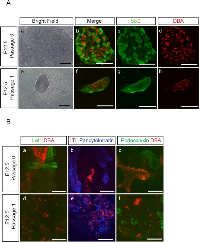
Fig 4. Suboptimal NPC maintenance in P1 aggregates.

(A) Aggregates were reconstituted from E12.5 embryonic kidneys and cultured for 7 days. Some of the E12.5 P0 aggregates were further passaged at day 7 to reconstitute P1 aggregates and cultured for another 7 days. The bright field images and the immune-staining for NPC marker, Six2 (green), and UB marker, DBA (red), of representative P0 (a-d) and P1 (e-h) aggregates show that P1 aggregates were smaller in size and contained fewer numbers of Six2+ NPC as compared to P0 aggregates. In contrast to the organized UB branching structure in P0 aggregates (b, d), UB cells in P1 (f, h) aggregates formed randomly scattered structures. (Scale bar = 500 μm). (B) E12.5 P0 and P1 aggregates were immuno-stained for: (a,d) renal vesicle marker, Lef1 (green)/UB marker, DBA (red); (b,e) proximal tubule marker, LTL (red)/epithelial marker, pancytokeratin (blue); (c,f) podocyte marker, podocalyxin (green)/UB marker, DBA (red), and show the development of Lef+-renal vesicle like structures (a), LTL+- (b,e) and podocalyxin+-(c,f) epithelial structures. (Scale bar = 100 μm)
