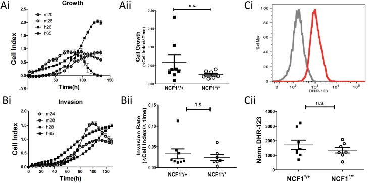
Fig 6. In vitro characteristics of NCF1*/* and NCF1*/+ MCA induced tumors.
Tumors cell lines were generated from MCA induced tumors by mechanical dissociation followed by treatment with Collagenase IV and Trypsin. Tumor cells were plated at low confluence on Xcelleigence E-plates and (Ai) growth was monitored over time and (Aii) cell growth rate was determined based on the logarithmic growth phase. (Bi) Tumor cells were seeded onto CIM-16 xCELLigence plates with serum medium gradient separated by matrigel plug to evaluate invasion for which the (Bii) invasion rate was compared between NCF1*/* and NCF1*/+ cell lines. (C) Tumor cells were labeled with DHR-123 and analyzed by flow cytometry.

