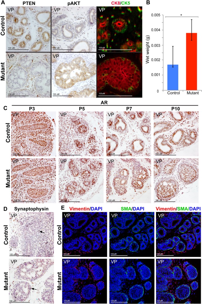
Fig 2. Nkx3.1:Cre;Pten Fl/Fl mice exhibit a cellular phenotype at P10.
(A) Antibody staining of P10 prostate sections demonstrates that mutant mice exhibit loss of PTEN, increased expression of pAKT, of organization of CK8 (red) luminal cells that fill the duct and CK5 (green) basal cells. (B) P10 mutant mice exhibit a statistically significant (*, p = 0.002, n = 10) increase in prostate wet weight compared to control mice. Antibody staining on sections of prostates demonstrates that (C) AR expression is similar in the mutant compared to controls at P3, P5, P7 and P10, (D) rare neuroendocrine cells marked by Synaptophysin are present at P10 in the mutant and controls (black arrow), and (E) Vimentin and Smooth Muscle Actin (SMA) expression shows that mesenchymal differentiation has taken place at P10 in the mutant similar to controls. Scale bars are as indicated. All sections are ventral prostate (VP).

