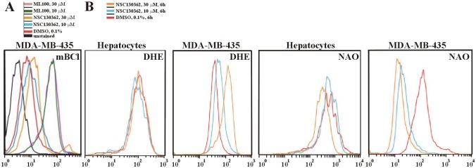
Fig 7.

(A) MDA-MB-435 cells that survived after NSC130362 treatment had elevated levels of GSH. Subconfluent MDA-MB-435 cells in a 6-well plate were treated for 6 h with NSC130362 (10 and 30 μM), or DMSO followed by staining with mBCl (40 μM) for 10 min and subjected to subsequent flow cytometry analysis. Mean fluorescence intensity was: 2.55 (unstained cells), 6.47 (DMSO-treated cells), 8.60 (10 μM NSC130362-treated cells), 11.60 (30 μM NSC130362-treated cells), 38.30 (10 μM ML100-treated cells), 37.30 (30 μM ML100-treated cells). (B) NSC130362 induced ROS generation and peroxidation of mitochondrial membrane lipid. Subconfluent MDA-MB-435 cells in a 6-well plate were treated for 6 h with either NSC130362 (10 and 30 μM) or DMSO followed by staining with DHE (10 μM) and NAO (5 nM) for 20 min and subjected to subsequent flow cytometry analysis.
