Figure 2.

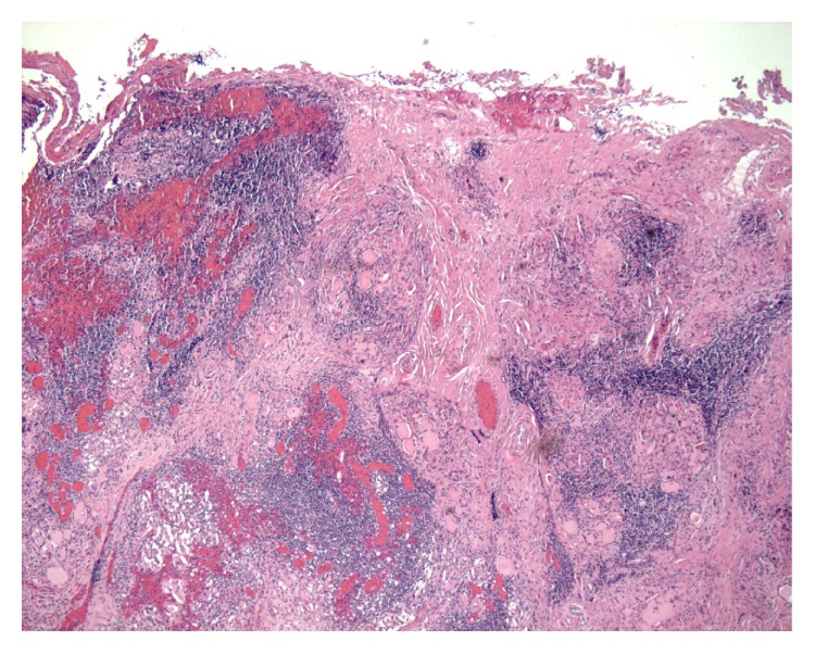
Case 2. Rare atrophic follicles with intraluminal colloid. Marked inflammation with extensive lymphocytic infiltration with some germinal centres and associated peripheral fibrous septa.

Case 2. Rare atrophic follicles with intraluminal colloid. Marked inflammation with extensive lymphocytic infiltration with some germinal centres and associated peripheral fibrous septa.