Figure 3.

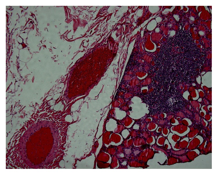
Case 3. Subcapsular nodular lymphocytic infiltration, medium-large size thyroid follicles with intraluminal colloid. On the left side perithyroidal adipose tissue with congested vessels.

Case 3. Subcapsular nodular lymphocytic infiltration, medium-large size thyroid follicles with intraluminal colloid. On the left side perithyroidal adipose tissue with congested vessels.