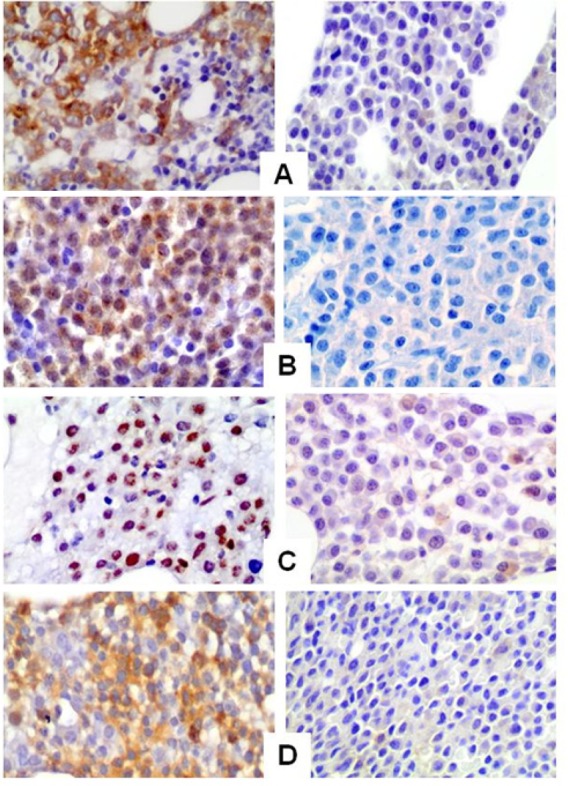
Figure 6. Representative immunohistochemical staining of MM bone marrow samples for p-AKT (A), p-mTOR (B), p-P706SK (C) and p-4E-BP1 (D). The right panel shows negative immunoreactivity, while the left panel shows strong positive immunoreactivity for the specific antibodies.

Original magnification 200x.
