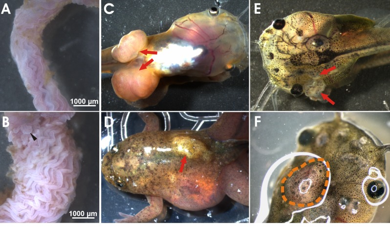
Figure 2. Neoplastic phenotypes observed in apc TALEN mRNA injected tadpoles and frogs.
Small intestine, cut open longitudinally, of wild type (WT) (A) and apc TALEN mRNA (40 pg) injected (B) adult frogs. In WT frogs, the epithelial lining of the duodenum is organized in parallel longitudinal folds. The intestines of apc TALEN injected animals are largely expanded and the intestinal folds are irregular and excessively undulated. Aberrant local protuberances are visible (black arrowhead). (C) Large fast growing tumors at the position where the hindlimbs emerge (red arrows). (D) Desmoid tumor, visible as a subcutaneous light colored mass (red arrow). (E) Epidermoid cysts (red arrows) originating at various positions in the tadpole skin. (F) Medulloblastoma visible as a large mass in the tadpole brain region.

