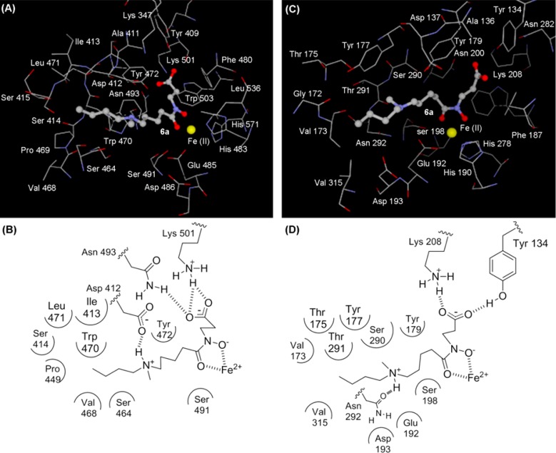
Figure 2.
(A) View of the conformation of compound 6a (ball and stick) docked into the JARID1A active site. (B) Schematic diagram of binding of compound 6a to JARID1A (a homology model based on the crystal structure of JMJD2A). (C) View of the conformation of compound 6a (ball and stick) docked into the JMJD2C active site (PDB code 2MXL). (D) Schematic diagram of binding of compound 6a to JMJD2C.

