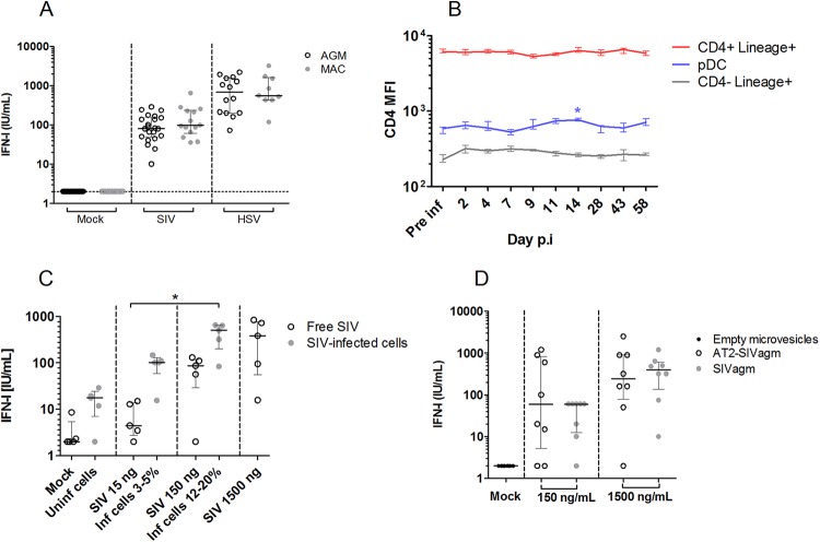
FIG 3.
SIV sensing capacity of AGM pDC. (A) SIVagm.sab92018 was used to stimulate AGM (n = 22) and SIVmac251 was used to stimulate Chinese rhesus MAC (n = 14) PBMCs. Alternatively, PBMCs were stimulated with HSV-1. (B) CD4 MFI was followed throughout SIVagm infection of four AGMs. Median and interquartile ranges are shown for CD4+ Lineage+ cells, pDC, and CD4− Lineage+ cells. Pre inf, preinfection time point; Day p.i, day postinfection. (C) SIVagm-infected SupT1 cells and free virions were used to stimulate AGM PBMCs (n = 5). Inf, infected; Uninf, uninfected. (D) SIVagm or AT2-inactivated SIVagm was used to stimulate AGM PBMCs (n = 8). Individual symbols represent distinct animals. Medians and interquartile ranges are shown. Vertical dashes separate the different viral preparations used to test pDC sensing. *, Friedman, P < 0.05.

