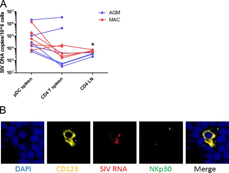
FIG 5.
In vivo pDC infection. (A) pDC and CD4+ T cells of chronically SIV-infected AGM (n = 7) and Chinese rhesus MAC (n = 5) were sorted from 2 × 10⁸ to 4 × 10⁸ splenocytes, yielding a median of 6,000 pDC after two subsequent sorts (purity, 91%). CD4+ T cells were also purified from lymph node (LN) cells (purity, 97%). SIV DNA was normalized to CCR5 and is represented as copies per million cells. Symbols represent individual animals. CD4+ T cells in LNs were infected to a higher extent in MAC than in AGM (*, Mann-Whitney, P = 0.016). SIV DNA copy numbers of spleen and LN CD4+ T cells were similar in the two species. (B) Fluorescence microscopy was performed on a LN of one chronically infected AGM. DAPI (blue) staining shows nuclei, CD123 (yellow) is expressed on pDC, SIV RNA (red) shows infected cells, and NKp30 (green) is a marker not expressed on pDC. The merge shows an overlap of CD123 and SIV signals.

