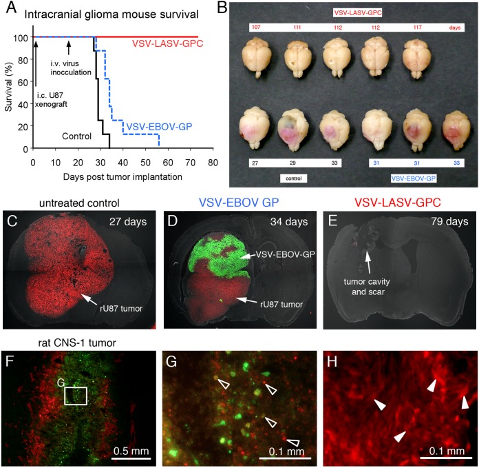
FIG 4.
Intravenous VSV-LASV-GPC destroys brain glioma and prolongs life indefinitely. CB17 SCID mice with unilateral striatal xenografts of human RFP-expressing rU87 glioma were treated with a single intravenous injection of either VSV-LASV-GPC, VSV-EBOV-GP (each 100 μl of 106 PFU), or saline (control) 15 days after tumor placement. (A) Kaplan-Meier curve showing modest survival benefit of VSV-EBOV-GP-treated mice compared to untreated control (median survival of 29 versus 34 days; n = 8 each group). In contrast, VSV-LASV-GPC-treated mice showed complete survival throughout the observation period (80 days, n = 8). Histological analysis showed no residual viable tumor mass, only scar tissue and tumor cavity (E). (B) The panel displays representative brains for each group. Brains from untreated control mice showed massive expansion of the tumor mass (pink), causing a significant midline shift of the longitudinal cerebral fissure. Large tumor mass and midline shift is also seen in brains from mice treated with VSV-EBOV-GP, indicating limited therapeutic effect. In contrast, brains from VSV-LASV-GPC-treated mice (top row) showed no visible expansion of the brain at the end of the observation period. (C) In control mouse, red glioma expanded substantially. (D) Mouse treated with VSV-EBOV-GP showed selective infection in tumor, but the infection was not complete. (E) Intravenous VSV-LASV-GPC completely eliminated tumor by 79 days. The majority of VSV-LASV-GPC-treated brains showed an empty tumor cavity, indicating successful oncolysis. (F) Syngeneic rat brain tumor model was established by stereotactically grafting RFP-expressing CNS-1 rat glioma into Lewis rats into the right striatum. VSV-LASV-GPC was applied to the area of the tumor 7 days after tumor placement; rats were euthanized 3 days later. VSV-LASV-GPC caused widespread oncolysis in the central portions of CNS-1 tumors with dead or dying cells, resulting in fading of the RFP and GFP signals and a morphological shift to cell debris (open arrowheads in panel G) from the whole-cell architecture seen in noninfected CNS-1 glioma (white arrowheads in panel H). Infection was restricted to the tumor. These data support the view that VSV-LASV-GPC not only targets human glioma but can also selectively infect rodent glioma within the rodent brain.

