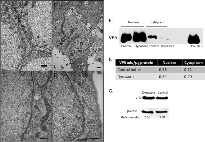
FIG 8.
Viral capsids are restricted to the nucleus in dynasore-treated cells. (A to D) SK-N-SH cells were infected with HSV-2(G) (MOI, 5 PFU/cell) for 1 h at 37°C. After unbound virus was washed off, the cells were overlaid with either a control medium containing 0.25% DMSO or a medium containing 80 μM dynasore. At 16 h p.i., cells were fixed and were prepared for electron microscopy. Representative images from control (magnification, ×10,000) (A and B) and dynasore-treated (magnification, ×20,000) (C and D) cells from two independent experiments are shown. Bars, 500 nm. (E) SK-N-SH cells were infected at an MOI of 10 PFU/cell, and 6 h p.i., dynasore or 0.25% DMSO in serum-free medium was added. The cells were harvested 18 h p.i.; cytoplasmic and nuclear fractions were prepared; and capsids were purified on 20- to 50% (wt/vol) sucrose gradients. The capsids were analyzed by Western blotting and were probed with an antibody to VP5. Purified HSV-2(G) was included as a positive control. (F) Blots were scanned, and relative optical density units (odu) normalized for total protein per lane are shown. Results are representative of 3 independent experiments. (G) Unfractionated infected SK-N-SH cell pellets were analyzed in parallel for VP5; representative results are shown. Blots were scanned, and VP5 odu relative to β-actin odu are shown below each lane.

