FIG 9.
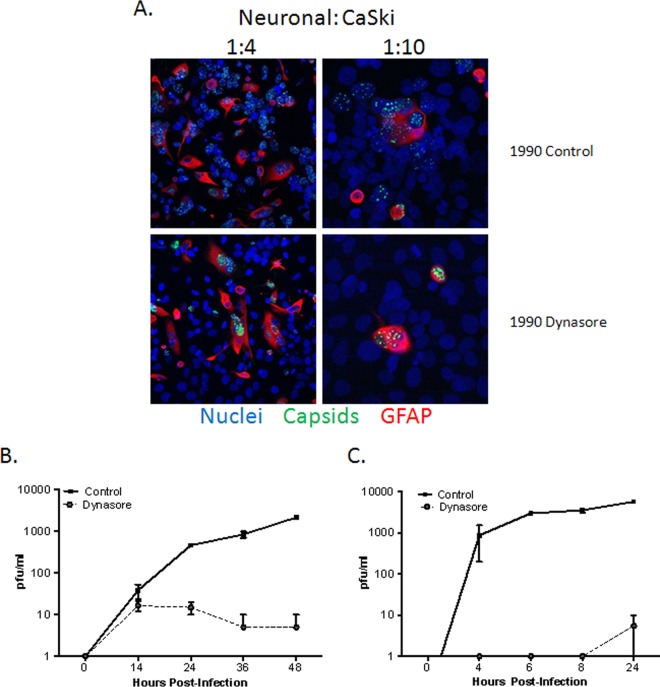
Dynasore prevents cell-to-cell spread. (A) Primary human neuronal cells were infected with HSV-1(KVP26GFP) (10 PFU/cell) for 1 h at 37°C. Then unbound virus was washed off, and cells were trypsinized and cocultured on a layer of untreated CaSki cells (∼ 90% confluence) on glass coverslips at a ratio of ∼1:4 or 1:10 (infected neuronal cells to uninfected CaSki cells). At 5 h postinfection, dynasore (80 μM in 0.25% DMSO) or a control medium (0.25% DMSO) was added to these cocultures. At 18 h p.i., cells were fixed with 4% paraformaldehyde and were stained with a primary anti-GFAP antibody (red); nuclei were stained with DAPI (blue). Images are representative of 3 independent experiments. 1990, 199V medium supplemented with 0.2% pooled human immunoglobulin (Sigma). (B) One-step growth of HSV-2 was quantified by infecting SK-N-SH cells with an MOI of 5 PFU/cell and adding dynasore or a control buffer to the overlay medium. The culture supernatants were harvested at the indicated times and were diluted 1:10 to decrease any residual dynasore to noninhibitory levels of ≤8 μM. Viral growth was quantified by determining titers on Vero cells. (C) To assess multistep growth, SK-N-SH cells were infected with an MOI of 0.02 PFU/cell, and dynasore or a control buffer was added to the overlay medium 6 h p.i. Culture supernatants were harvested at the indicated times, and titers were determined on Vero cells as for the single-step growth curve. Data shown are from two experiments conducted in duplicate; growth curves are significantly different (P < 0.001) by two-way analysis of variance.

