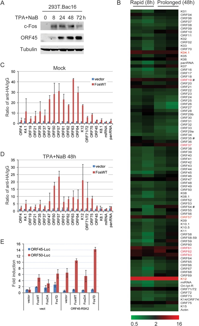
FIG 5.
Genome-wide screening for c-Fos binding to KSHV promoters. (A and B) Bac16-harboring 293T cells were induced with 20 ng/ml TPA and 0.3 mM NaB. (A) c-Fos accumulation was confirmed by Western blotting as indicated. (B) At 8 h or 48 h, the cells were collected and fixed. After sonication and preclearing, the same amount of cellular DNA lysates was subjected to a ChIP assay with an anti-c-Fos antibody, with normal IgG used as the negative control. The whole panel of cellular and viral DNAs was extracted and analyzed by real-time PCR in triplicate in two independent experiments, the relative DNA levels were normalized to control IgG, and the relative fold enrichment values were calculated relative to that of β-actin promoters. The results are shown as a heat map. #, rapid (8 h) versus prolonged (48 h), P < 0.05. The positive c-Fos binding under both conditions is indicated in red. (C and D) Control or HA-c-Fos constructs were transfected into Bac16-harboring 293T cells for 48 h, and then the cells were left uninduced (C) or induced as described above for 48 h (D). ChIP assays were performed with an anti-HA antibody, and viral DNAs were analyzed by real-time PCR in triplicate in two independent experiments. (E) Control, wild-type c-Fos (FosWT), constitutively active c-Fos (FosTD), or double phosphorylation site-mutated c-Fos (FosDA) constructs were cotransfected into HEK293 cells with ORF45-luc and ORF50-luc promoter reporters in the absence or presence of ORF45 and RSK2 overexpression. Thirty-six hours later, the dual luciferase activity was measured, and the data are shown as the means from three independent experiments performed in duplicate.

