FIG 2.

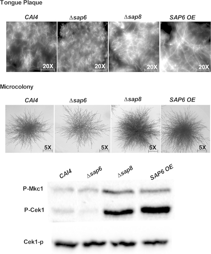
Architectures of in vivo fungal plaques and microcolony formation of the Δsap8 and SAP6OE strains show similar higher density and elongated filaments. (Top) Fungal plaques were removed from tongues, stained with calcofluor white, and observed under a DAPI (4′,6-diamidino-2-phenylindole) filter at ×20 magnification. Plaques from tongues infected with the Δsap8 and SAP6 OE strains contained fungal cells with longer and more compact hyphae than plaques from tongues infected with the Δsap6 and WT CAI4 strains. (Middle) Microcolonies of the Δsap8 and SAP6 OE strains grown under 5% CO2 were denser and more highly filamentous than those of the CAI4 strain, while the Δsap6 strain had the thinnest microcolony formation, although filament length was similar to that of the WT. (Bottom) Higher levels of Cek1 phosphorylation were found in fungal cells from tongue plaques infected with the Δsap8 and SAP6 OE strains than in tongue plaques infected with the CAI4 and Δsap6 strains.
